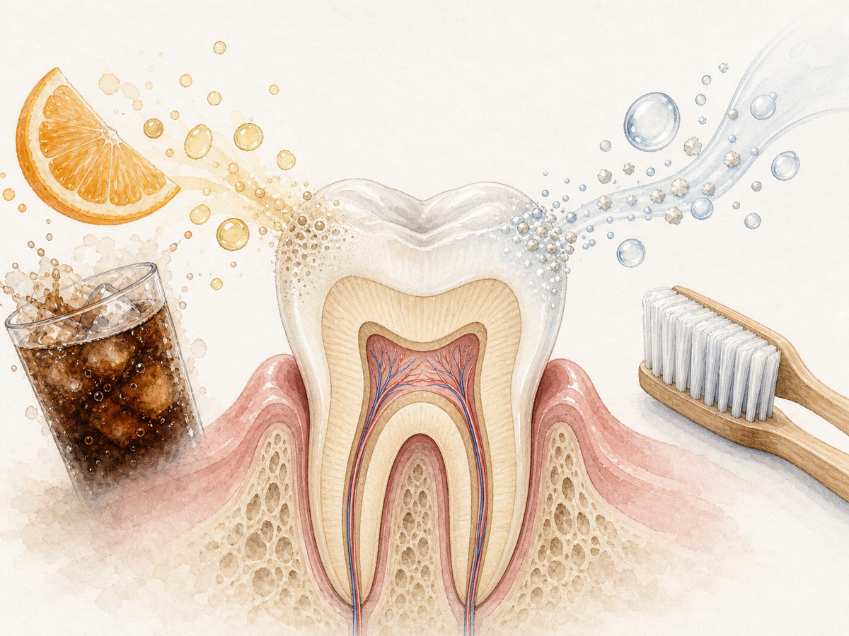









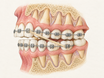


The alveolar process is the ridge of bone in your upper and lower jaws that forms the sockets around your teeth. It works with the tooth-supporting ligament to keep teeth in place and absorb chewing forces. When this bone shrinks or breaks down, teeth can loosen, and common dental treatments such as implants and dentures become harder to plan. Protecting it means controlling plaque, treating gum disease early, and asking about bone preservation before a tooth is removed.
Practical rule: If you need a tooth removed, ask your dentist whether ridge preservation is appropriate. This can limit the bone shrinkage that often follows an extraction.
Where It Sits in the Jaw
The alveolar process is part of the maxilla (upper jaw) and mandible (lower jaw). It is not the whole jawbone. It is the curved, tooth-bearing ridge that sits on top of the deeper basal bone — the main body of the jawbone — and surrounds the roots of the teeth. Anatomical guidance defines it as the part of the upper and lower jaws that forms and supports the tooth sockets.
Think of it like this: the upper ridge sits above your teeth, and the lower ridge sits below them. The top rim of this ridge is the alveolar crest. The crest is the highest edge of the bone between teeth.
The alveolar process only develops after teeth form and push through the gums. It is a tooth-dependent structure, not the main body of the jaw. It develops along with the teeth and can resorb after teeth are removed, according to alveolar bone science.
The Layers That Make It Work
The alveolar process is built in layers so it can be both strong and adaptable.
- The outer cortical plate is a compact, dense layer of bone.
- Inside is trabecular bone, a spongy, lattice-like bone with small spaces.
- Lining each tooth socket is a thinner layer called alveolar bone proper.
This combination matters. The dense outer layer resists bending during biting, while the spongy inner layer absorbs and spreads forces. The socket-lining bone is also called the cribriform plate, or “sieve-like plate,” because it contains many small canals. On dental X-rays, that same lining often appears as a thin, dense white line called the lamina dura. This layered design is a central part of alveolar bone structure and function.
The Socket, Lamina Dura, and Bundle Bone
Each tooth sits in a socket called an alveolus. The socket is not just a smooth hole. Its inner wall contains bundle bone, which is the specialized bone where the tooth-supporting fibers of the periodontal ligament insert. Those anchoring fibers are sometimes called Sharpey’s fibers. A review of alveolar bone describes this socket wall as the bone lining the alveolus and providing attachment for the ligament.
On an X-ray, the socket wall appears as the lamina dura, the thin dense line that outlines the root. Dentists use this line as a landmark because a healthy socket wall usually looks crisp and continuous. The socket, bundle bone, and lamina dura work together to create a precise attachment for each individual tooth, rather than a generic bone surface.
How It Anchors Teeth
Teeth are not fused directly into the jaw. They are suspended by the periodontal ligament, a tough band of tissue between the root and bone. Collagen fibers from this ligament run from cementum, the thin mineralized coating on the root, into the alveolar bone proper. This arrangement holds the tooth in its socket while also allowing slight, normal movement.
That design has two jobs. First, it spreads chewing forces across a wider area of bone instead of concentrating them in one spot. Second, it lets the tooth give a little under pressure, like a shock absorber. The alveolar process is the bone half of this suspension system. Biomechanical research supports the idea that the bone-periodontal ligament-tooth complex distributes and dampens chewing forces while permitting controlled tooth displacement.
The Alveolar Crest and Interdental Septa
The alveolar crest is the highest rim of bone between teeth. The interdental septa are the bony walls that separate one socket from the next. Their height and shape directly affect how much support a tooth has.
Dental exams track these landmarks because they are sensitive to early damage. In gum disease, bone often shrinks at the crest first. X-rays help detect this. Clinical guidance on dental radiographs notes that intraoral X-rays have moderate to high sensitivity for assessing alveolar bone loss in periodontitis.
On a healthy bitewing X-ray, the distance from the place where the crown meets the root to the alveolar crest is normally small. One study on alveolar crest level found that a radiographic distance of 0.4 to 1.9 mm is consistent with no crestal bone loss. A widening gap can be an early sign of periodontal disease.
Why It Remodels and Shrinks
Alveolar bone is highly responsive to mechanical load, meaning the physical stress of biting and chewing. That responsiveness is why teeth can be moved and also why bone shrinks after teeth are lost. The bone loss side of this process is called resorption.
Common triggers for remodeling or shrinkage include:
- Orthodontic movement from braces.
- Tooth drift or shifting after a nearby tooth is lost.
- Tooth extraction.
- Edentulism, or the loss of all teeth.
- Changes in bite forces from missing or damaged teeth.
When a tooth is removed and the socket heals on its own, the ridge can lose both width and height. A systematic review and meta-analysis concluded that variable amounts of alveolar bone resorption occur after unassisted socket healing, depending on tooth type. That is why a socket can look sunken or narrow after extraction.
Why It Matters Clinically
Alveolar bone volume and quality affect nearly every area of dental care.
- Gum and bone health: Periodontal support depends on how much ridge bone remains.
- Implant placement: A dental implant needs enough bone height and width to stabilize it.
- Denture fit: Dentures rest on the ridge, so a well-preserved ridge helps them stay in place.
- Extractions: How the socket heals affects future options for replacement.
- Orthodontics: Braces rely on the bone’s ability to remodel around moving roots.
Preserving alveolar bone is now considered important in implant-focused rehabilitation, according to a chapter on alveolar bone science. Bone volume also tends to be lower in atrophic, or shrunken, sites, as reported in a systematic review of alveolar bone architecture.
What Damages It and How to Protect It
The biggest direct threat is periodontitis, a severe form of gum disease that destroys the tooth-supporting tissues, including alveolar bone. The EFP S3 clinical practice guideline defines periodontitis as progressive destruction of the tooth-supporting apparatus. Other potential threats include trauma to the teeth or jaw, smoking, uncontrolled diabetes, and poorly fitting false teeth or dental appliances that place uneven pressure on the ridge.
Clinical guidelines emphasize that smoking and diabetes are important risk factors for periodontitis, and that controlling them is part of protecting the tooth-supporting tissues, including alveolar bone (EFP S3 clinical practice guideline).
You can protect alveolar bone with practical daily habits:
- Clean plaque away daily by brushing and cleaning between the teeth.
- Keep regular dental visits so gum disease and bone loss can be found early.
- Treat gum disease early instead of waiting until teeth are loose.
- If you smoke, seek support to quit.
- If you have diabetes, work with your medical and dental team to keep blood sugar controlled.
- If a tooth must be removed, ask about ridge preservation, a treatment designed to limit bone shrinkage after extraction.
Ridge preservation has been shown to reduce horizontal and vertical bone loss compared with leaving a socket to heal empty, according to a systematic review and meta-analysis.
See a dentist if you notice loose teeth, receding gums, bleeding when cleaning, a persistent bad taste, or a tooth that feels like it has shifted. These symptoms can be signs of gum disease or bone loss, but they need a professional evaluation to understand the cause.
Alveolar Process vs Basal Bone
It is easy to confuse the alveolar process with the whole jawbone, but the difference matters.
| Feature | Alveolar process | Basal bone |
|---|---|---|
| Main role | Holds teeth in their sockets | Forms the main body of the jaw |
| Depends on teeth? | Yes, strongly | No |
| After tooth loss | Can shrink over time | Generally remains |
| Dental relevance | Gum disease, ridge preservation, implants, dentures | Anchorage for some implants when the ridge is thin |
The alveolar process is tooth-dependent. The deeper basal bone is the structural base of the jaw that remains even when the ridge shrinks. This distinction is why some implant approaches use basal bone when severe alveolar ridge loss makes conventional implants difficult. One case report describes basal bone as denser and less likely to resorb than alveolar bone (basal implants for the edentulous mandible).
Keep the tooth-bearing ridge healthy while teeth are present. If a tooth is lost, talk to your dentist early about options to preserve the bone before it shrinks.
 Best Overall
 Best Value
 Best for Sensitive Teeth
This article has been reviewed by an oral health professional for accuracy. It is intended to provide general educational information and should not be used as a substitute for personalized medical advice, diagnosis, or treatment. If you have questions or concerns about your oral health, please consult a dentist, physician, or other qualified healthcare provider.